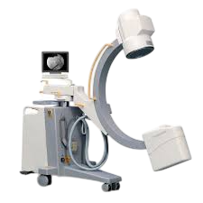
In the fast-evolving world of healthcare, diagnostic imaging has made significant strides in recent years, offering medical professionals better tools for diagnosing and treating a wide range of conditions. As technology advances, the capabilities of imaging devices are expanding, providing clearer images, faster results, and even more non-invasive procedures. In this blog, we explore some of the latest innovations in diagnostic imaging technology that are transforming the medical field.
1. Artificial Intelligence (AI) Integration in Imaging
One of the most exciting developments in diagnostic imaging is the integration of artificial intelligence (AI). AI-powered imaging systems can analyze vast amounts of data quickly and accurately, helping radiologists detect patterns that might be missed by the human eye.
AI in Imaging Workflow:
- Automatic Image Analysis: AI can assist in interpreting images from MRIs, CT scans, and X-rays, identifying abnormalities such as tumors, fractures, and lesions with precision.
- Workflow Optimization: AI algorithms streamline the imaging process by automating routine tasks like sorting images and generating reports, reducing the workload on medical staff and improving the speed of diagnosis.
- Enhanced Diagnostic Accuracy: With machine learning models trained on millions of cases, AI has shown remarkable accuracy in diagnosing certain conditions, such as lung cancer and brain tumors, even in early stages.
2. Portable Imaging Devices
Another major innovation in diagnostic imaging is the rise of portable imaging devices. Traditionally, diagnostic imaging required bulky, expensive equipment that was only available in hospitals or large clinics. Today, compact, portable devices allow healthcare providers to deliver diagnostic imaging services in almost any setting.
Key Benefits of Portable Imaging:
- Accessibility in Remote Areas: Portable devices, such as handheld ultrasound machines and portable X-ray units, enable healthcare delivery in remote or underserved areas where access to full-scale diagnostic imaging is limited.
- Emergency Use: In critical care situations, portable devices allow for real-time diagnostic imaging in emergency rooms or even at the patient’s bedside, providing immediate insights to guide treatment decisions.
- Cost-Effectiveness: Portable devices are often more affordable than their full-size counterparts, making them an attractive option for smaller clinics and practices.
3. 3D and 4D Imaging
3D imaging has been around for some time, but recent advancements are pushing the boundaries of what’s possible. 4D imaging, which adds the dimension of time to 3D scans, allows medical professionals to visualize how organs and structures move in real-time.
Applications of 3D and 4D Imaging:
- Fetal Monitoring: 3D and 4D ultrasound imaging provide detailed images of a fetus in the womb, allowing doctors to monitor development and detect any abnormalities early on.
- Surgical Planning: Surgeons can now use 3D imaging to create detailed models of organs and tissues, enabling them to plan procedures with greater precision, reducing risks and improving outcomes.
- Tumor Tracking: 4D imaging is particularly useful for tracking the movement of tumors, especially in regions like the lungs, where breathing causes constant shifts. This real-time visualization helps in targeting tumors more effectively during radiation therapy.
4. Photon-Counting CT Scanners
Photon-counting CT (PCCT) scanners represent a breakthrough in computed tomography technology. Unlike traditional CT scanners that measure the total energy of X-rays that pass through the body, photon-counting scanners detect individual X-ray photons, providing a more detailed and accurate picture.
Advantages of Photon-Counting CT:
- Sharper Image Quality: PCCT provides higher resolution images with less noise, which is crucial for detecting small lesions or subtle abnormalities.
- Lower Radiation Dose: Since photon-counting detectors are more sensitive, they require less radiation to produce clear images, making this technology safer for patients.
- Multi-Energy Imaging: PCCT can differentiate between different materials (such as bone, tissue, and blood) more effectively, which is useful for a variety of diagnostic applications, including cardiovascular imaging.
5. Hybrid Imaging Systems
Hybrid imaging systems combine multiple imaging modalities into one machine, offering a more comprehensive view of the body’s structures and functions. PET/CT (Positron Emission Tomography/Computed Tomography) and PET/MRI systems are prime examples of this innovation.
Benefits of Hybrid Imaging:
- Improved Diagnostic Accuracy: By combining functional imaging (PET) with anatomical imaging (CT or MRI), hybrid systems provide a more complete picture of the patient’s condition. For example, PET can detect metabolic changes in tissues, while CT or MRI provides detailed structural images.
- Faster Diagnoses: Hybrid imaging eliminates the need for multiple separate scans, reducing the time to diagnosis and allowing for quicker treatment decisions.
- Cancer Detection and Monitoring: PET/CT and PET/MRI are especially valuable in oncology, where they can detect cancer, assess the effectiveness of treatments, and monitor for recurrence.
6. Wearable Imaging Technology
While still in the early stages of development, wearable imaging devices are set to revolutionize how diagnostic imaging is performed. These devices can be worn by patients, providing continuous monitoring and real-time imaging without the need for bulky, stationary equipment.
Examples of Wearable Imaging:
- Wearable Ultrasound: Some researchers are developing flexible, wearable ultrasound patches that can be applied to the skin to monitor internal organs continuously over time.
- MRI Gloves: Experimental MRI gloves allow for detailed imaging of hand anatomy in motion, which could be useful for diagnosing conditions like arthritis or injuries in athletes.
7. Spectral Imaging
Spectral imaging is an advanced technique that captures multiple wavelengths of light or energy to provide more detailed information than traditional imaging methods. This is especially useful in CT imaging, where spectral CT can distinguish between different tissue types, enhancing the contrast in the images.
Advantages of Spectral Imaging:
- Improved Tissue Characterization: Spectral imaging provides more detailed information about the composition of tissues, which is particularly useful for identifying specific types of lesions or differentiating between benign and malignant tumors.
- Better Contrast in Low-Dose Imaging: Spectral CT can produce high-contrast images even with lower radiation doses, making it safer for patients.
Conclusion
The latest innovations in diagnostic imaging technology are revolutionizing the way healthcare providers diagnose and treat their patients. From AI-powered imaging to portable devices and hybrid systems, these advancements are improving the accuracy, speed, and accessibility of medical imaging. As these technologies continue to evolve, the future of diagnostic imaging promises even more exciting developments, ultimately enhancing patient care and outcomes.
If your facility is looking to upgrade or invest in imaging equipment, it’s crucial to stay informed about these advancements to ensure you provide the best care possible. At Red Stone Medical, we specialize in helping medical practices find the right imaging equipment for their needs, whether it’s state-of-the-art new systems or high-quality refurbished machines. Contact us today to learn more about our offerings.

